Search results
Search for "X-ray diffraction" in Full Text gives 521 result(s) in Beilstein Journal of Nanotechnology. Showing first 200.
Photocatalytic degradation of methylene blue under visible light by cobalt ferrite nanoparticles/graphene quantum dots
Beilstein J. Nanotechnol. 2024, 15, 475–489, doi:10.3762/bjnano.15.43

- synthesize cobalt ferrite nanoparticles/graphene quantum dots (CF/GQDs). The material was prepared from a homogeneous mixture of iron nitrate, cobalt nitrate, and starch at 140, 180 and 200 °C in a 24 h thermal hydrolysis process. The obtained materials were characterised by using X-ray diffraction, scanning
- benzoquinone (C6H4O2) was provided by Sigma-Aldrich, USA. Sodium hydroxide (NaOH, ≥96%) and hydrogen chloride (HCl, 38%) were purchased from Xilong, China. Instruments X-ray diffraction (XRD) patterns were recorded by using a D8 Advance (Bruker, Germany) with Cu Kα radiation (λ = 0.154 nm). Fourier-transform


Controllable physicochemical properties of WOx thin films grown under glancing angle
Beilstein J. Nanotechnol. 2024, 15, 350–359, doi:10.3762/bjnano.15.31

- bias voltage was applied to the p-Si substrates, whereas the WOx films were kept grounded. The crystallinity of the WOx films was examined using X-ray diffraction (XRD) (Bruker) under Bragg–Brentano geometry (θ–2θ) in an angular window of 2θ = 20° to 80°. The chemical composition of the WOx films was


Vinorelbine-loaded multifunctional magnetic nanoparticles as anticancer drug delivery systems: synthesis, characterization, and in vitro release study
Beilstein J. Nanotechnol. 2024, 15, 256–269, doi:10.3762/bjnano.15.24

- -resolution analytical electron microscope (FE-SEM, Thermo Scientific, Apreo 2S LoVac) and a scanning transmission electron microscope (STEM, Phillips XL, 30 ESEM-FEG/EDAX) operating at 120 kV acceleration voltage. The structure of the nanoparticles was analyzed by X-ray diffraction (XRD, PANalytical, Xpert
- between peak broadening and particle size in X-ray analysis. In this equation, the symbols D, K, λ, β, and θ represent the particle size, Scherrer shape factor (here 0.89), X-ray wavelength (0.15418 nm), half-maximum width, and diffraction angle, respectively [43]. Using the X-ray diffraction (XRD
- × magnification. (d) Particle diameter distribution of Fe3O4 NPs. (a) X-ray diffraction patterns of Fe3O4 NPs. (b) FTIR spectra of Fe3O4 NPs. (c) Hysteresis loop for Fe3O4 NPs. FESEM images of (a) PDA/Fe3O4 NPs (1:1), (b) PDA/Fe3O4 NPs (2:1), and (c) PDA/Fe3O4 NPs (4:1), all 250,000× magnification. (d) FTIR


Modification of graphene oxide and its effect on properties of natural rubber/graphene oxide nanocomposites
Beilstein J. Nanotechnol. 2024, 15, 168–179, doi:10.3762/bjnano.15.16

- characterized by X-ray diffraction (XRD), Fourier-transform infrared spectroscopy, contact angle, thermal gravimetric analysis, and scanning electron microscopy. The XRD results showed the appearance of an amorphous region of silica particles at a diffraction angle of 22°. The formation of silica was
- conditions to determine the ideal condition to modify GO for grafting onto NR. The GO-VTES products were characterized using X-ray diffraction (XRD), contact angle, 29Si NMR, Fourier-transform infrared spectroscopy (FTIR), and morphology analysis. The GO-VTES was expected to improve the mechanical properties
- , 200 g of DPNR (DRC 20%, SDS 0.2%), and with the same amount of TEPA/TBHPO as in the preparation of DPNR/GO-VTES. Characterizations X-ray diffraction patterns of graphite and GO were acquired by using a Panalytical X'Pert Pro X-ray diffractometer. The measurements were performed at room temperature


Assessing phytotoxicity and tolerance levels of ZnO nanoparticles on Raphanus sativus: implications for widespread adoptions
Beilstein J. Nanotechnol. 2024, 15, 115–125, doi:10.3762/bjnano.15.11

- on Raphanus sativus (R. sativus) concerning its tolerance levels, toxicity, and accumulation. ZnO NPs were synthesized by the wet chemical method and characterized by powder X-ray diffraction (PXRD), Fourier-transform infrared (FTIR) spectroscopy, ultraviolet–visible (UV–vis) spectroscopy, dynamic
- synthesized ZnO NPs were characterized via several techniques such as powder X-ray diffraction (PXRD), Fourier-transform infrared (FTIR) spectroscopy, solid-UV–vis spectroscopy, dynamic light scattering (DLS), and scanning electron microscopy (SEM). Then the potential phytotoxicity of the synthesized ZnO NPs


Berberine-loaded polylactic acid nanofiber scaffold as a drug delivery system: The relationship between chemical characteristics, drug-release behavior, and antibacterial efficiency
Beilstein J. Nanotechnol. 2024, 15, 71–82, doi:10.3762/bjnano.15.7

- separation during the electrospinning process [17][38][39], leading to the formation of a BBR-rich phase on the surface of nanofibers. The crystallinity of the PLA pellet and electrospun nanofiber scaffolds were examined by X-ray diffraction (XRD) analysis (Figure 3B). The XRD pattern of the PLA pellet shows
- chemical characteristics of prepared scaffolds in the wavelength range of 200–3200 cm−1. X-ray diffraction measurements of PLA pellets and BBR-loaded PLA nanofiber scaffolds were analyzed with Cu Kα radiation in a 2θ range from 5 to 80° using EQUINOX 5000 – Thermo Scientific X-ray diffractometer. Static


Dual-heterodyne Kelvin probe force microscopy
Beilstein J. Nanotechnol. 2023, 14, 1068–1084, doi:10.3762/bjnano.14.88


Experimental investigation of usage of POE lubricants with Al2O3, graphene or CNT nanoparticles in a refrigeration compressor
Beilstein J. Nanotechnol. 2023, 14, 1041–1058, doi:10.3762/bjnano.14.86

- nanoparticles is presented separately in the subsequent sections to verify the catalog information provided by the manufacturer. In the characterization of the nanoparticles used in the study, field-emission scanning electron microscopy (FE-SEM), energy-dispersive X-ray spectroscopy (EDS), and X-ray diffraction
- micrograph and b) EDS analysis of Al2O3 nanoparticles. X-ray diffraction pattern of Al2O3 nanoparticles [21]. The a) FE-SEM micrograph with a scale bar of 20 µm, b) FE-SEM micrograph with a scale bar of 10 µm, and c) EDS analysis of the graphene nanoplatelets. The XRD pattern of the graphene nanoplatelets


Exploring internal structures and properties of terpolymer fibers via real-space characterizations
Beilstein J. Nanotechnol. 2023, 14, 1004–1017, doi:10.3762/bjnano.14.83

- , this distinction in fundamental chemistry has significant implications for the structures and properties of the resulting fibers. To date, structure–property characterizations of Technora® in the literature have primarily focused on (i) X-ray diffraction (XRD), (ii) nuclear magnetic resonance (NMR


A wearable nanoscale heart sound sensor based on P(VDF-TrFE)/ZnO/GR and its application in cardiac disease detection
Beilstein J. Nanotechnol. 2023, 14, 819–833, doi:10.3762/bjnano.14.67

- . Composition and β-phase content of the piezoelectric composite films were analyzed using X-ray diffraction. The morphology of the composite film fibers was observed through scanning electron microscopy. Finally, the P(VDF-TrFE)/ZnO/graphene composite film was encapsulated in a sandwich-structure heart sound
- underwent characterization through electron microscopy, X-ray diffraction (XRD), and piezoelectric performance testing. The results indicated that the piezoelectric film with a composition ratio of 12% P(VDF-TrFE) + 10% ZnO + 0.1% GR exhibited superior performance regarding various aspects. Consequently, in
- -TrFE)/ZnO flexible piezoelectric composite films without GR. Analysis of X-ray diffraction patterns indicated that the β-phase content of P(VDF-TrFE)/ZnO/GR piezoelectric composite films was higher than that of P(VDF-TrFE)/ZnO and P(VDF-TrFE). P(VDF-TrFE)/ZnO/GR was packaged into a flexible


Nanostructured lipid carriers containing benznidazole: physicochemical, biopharmaceutical and cellular in vitro studies
Beilstein J. Nanotechnol. 2023, 14, 804–818, doi:10.3762/bjnano.14.66

- medium observed after the initial stage could be attributed to the gradual release of drug molecules from the matrix core, where the drug is mainly located according to X-ray diffraction (XRD) results [33]. Remarkably, although our NLC possess a comparatively lower drug load, the maximal accumulated drug
- to the dispersion for contrast enhancement. X-ray diffraction structural analysis Small angle X-ray scattering/wide angle X-ray scattering measurements were performed using a XEUSS 2.0 equipment (XENOCS, France). Patterns were registered with two synchronous 2D photon-counting pixel X-ray detectors


Silver nanoparticles loaded on lactose/alginate: in situ synthesis, catalytic degradation, and pH-dependent antibacterial activity
Beilstein J. Nanotechnol. 2023, 14, 781–792, doi:10.3762/bjnano.14.64

- potential measurements, which were measured on a nanoPartica Horiba SZ-100 (Japan). Fourier-transform infrared (FTIR) spectra were obtained using a Bruker Tensor 27 FTIR spectrophotometer (Germany). X-ray diffraction (XRD) patterns were collected using a Bruker D8 Advance X-ray diffractometer. The


In situ magnesiothermic reduction synthesis of a Ge@C composite for high-performance lithium-ion batterie anodes
Beilstein J. Nanotechnol. 2023, 14, 751–761, doi:10.3762/bjnano.14.62

- and transferred to a ceramic crucible. The mixture was heated at 750 °C in argon gas flow for 3 h. The obtained solid was re-ground and denoted as Ge/C-SS750. Material characterization X-ray diffraction measurements (XRD, Bruker D8 Advance with Cu Kα radiation (λ = 1.5406 Å) at 40 kV and 40 mA) were


A graphene quantum dots–glassy carbon electrode-based electrochemical sensor for monitoring malathion
Beilstein J. Nanotechnol. 2023, 14, 701–710, doi:10.3762/bjnano.14.56

- hydrothermal process with glucose as a precursor undergoing carbonization. Different spectroscopic techniques were used to analyze the optical characteristics of GQDs, including UV–visible, photoluminescence, FTIR, and Raman spectroscopy. Atomic force microscopy, transmission electron microscopy, and X-ray
- diffraction were used to characterize the morphological and structural properties of GQDs. An electrochemical sensor was developed by drop casting GQDs on a glassy carbon electrode (GCE). The sensor detects the organophosphate pesticide malathion in a selective and sensitive manner. Using cyclic voltammetry


The microstrain-accompanied structural phase transition from h-MoO3 to α-MoO3 investigated by in situ X-ray diffraction
Beilstein J. Nanotechnol. 2023, 14, 692–700, doi:10.3762/bjnano.14.55

- Sciences, Beijing, 100190, China 10.3762/bjnano.14.55 Abstract In situ X-ray diffraction indicates that the structural phase transition from h-MoO3 to α-MoO3 is a first-order transition with a phase transition temperature range of 378.5–443.1 °C. The linear coefficients of thermal expansion of h-MoO3 are
- α-MoO3 is still unclear. Here, to reveal the features of the structural phase transition from h-MoO3 to α-MoO3, we performed in situ X-ray diffraction experiments at temperatures ranging from 30 to 450 °C. The Rietveld refinement results indicate water molecules at the (0 0 0.25) site inside the
- α-MoO3 To observe the crystal structure evolution of h-MoO3 induced by temperature, a thoroughly powdered sample was used to perform in situ X-ray diffraction measurements during heating from 30 to 450 °C, as shown in Figure 1a. At 30 °C, all diffraction peaks can be well indexed to the hexagonal


Titania nanoparticles for photocatalytic degradation of ethanol under simulated solar light
Beilstein J. Nanotechnol. 2023, 14, 616–630, doi:10.3762/bjnano.14.51

- in the second batch of samples, that is, in series “b”. The elemental composition of the TiO2 powders was estimated by EDS performed inside a scanning electron microscope, FEI Quanta Inspect S, at 15 kV in high vacuum. The crystalline structures and phase concentrations were determined from X-ray
- diffraction (XRD) patterns, measured by an X-ray diffractometer Panalytical X’Pert MPD theta–theta, and the morphological properties were determined by transmission electron microscopy (TEM), high-resolution transmission electron microscopy (HRTEM), and selected-area electron diffraction (SAED) measurements


Mixed oxides with corundum-type structure obtained from recycling can seals as paint pigments: color stability
Beilstein J. Nanotechnol. 2023, 14, 467–477, doi:10.3762/bjnano.14.37

- colorimetric stability of the aluminates applied as synthetic inorganic pigments [14]. Characterization The samples were characterized by powder X-ray diffraction (XRD) carried out in a Bruker D2 Phaser Diffractometer (Berlin, Germany) with Cu Kα emission (λ = 1.5418 Å) and equipped with a LynxEye high


Evaluation of electrosynthesized reduced graphene oxide–Ni/Fe/Co-based (oxy)hydroxide catalysts towards the oxygen evolution reaction
Beilstein J. Nanotechnol. 2023, 14, 420–433, doi:10.3762/bjnano.14.34
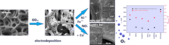
- probably inhibited the electrodeposition process of NiFe and CoNiFe on its surface. This may be the reason for the slower stabilization of the synthesis current density observed in the chronoamperograms (Figure 1a). X-ray diffraction, X-ray photoemission spectroscopy and X-ray absorption spectroscopy
- ]. The spectra were obtained using the total electron yield (TEY) detection mode, which can sample down to a depth of a few nanometers at room temperature. The beamline optics was optimized to perform the experiment with an energy resolution of 200 meV and better. X-ray diffraction (XRD) measurements


Formation of nanoflowers: Au and Ni silicide cores surrounded by SiOx branches
Beilstein J. Nanotechnol. 2023, 14, 133–140, doi:10.3762/bjnano.14.14

- atomic numbers show brighter contrasts. EDS measurements were performed to obtain the element distribution in the target areas. X-ray diffraction (XRD, Siemens D-5000) analyses were conducted in Bragg–Brentano mode using Cu Kα irradiation at 40 kV. The height distribution of the areas of interest was


Liquid phase exfoliation of talc: effect of the medium on flake size and shape
Beilstein J. Nanotechnol. 2023, 14, 68–78, doi:10.3762/bjnano.14.8

- powder was exfoliated in each liquid medium by exposure to mechanical energy provided by an ultrasonic bath (full details in the Experimental section). Talc was manually milled down to a fine powder and characterized by X-ray diffraction (XRD). Figure 1a displays the results. All peaks are assigned to
- obtaining information on thousands of flakes and using appropriate statistical descriptions to analyze the data. Experimental Materials. Talc was obtained through a donation of a sample from Minas Gerais state, Brazil. X-ray diffraction (XRD) was performed to characterize the sample composition. The rock
- measurements were performed on silicon substrates with an oxide layer, Si/SiOx. Substrates were functionalized with (3-aminopropyl)triethoxysilane (APTES) following the procedure reported by Fernandes and co-workers [24]. X-ray diffraction. XRD was performed in a Rigaku Geigerflex 2037 diffractometer with a


Two-step single-reactor synthesis of oleic acid- or undecylenic acid-stabilized magnetic nanoparticles by thermal decomposition
Beilstein J. Nanotechnol. 2023, 14, 11–22, doi:10.3762/bjnano.14.2

- , and composition via several techniques, such as transmission electron microscopy, dynamic light scattering, thermogravimetric analysis, Fourier-transform infrared spectroscopy/attenuated total reflectance, 57Fe Mössbauer spectroscopy, and X-ray diffraction. The effect of unsaturated oleic (OA) and
- were uniform and the single spots were not visible proved that the crystallites were very small. These results correspond well with data from X-ray diffraction (XRD), according to which the average size of the crystallites for all prepared nanoparticles was 4.5–9 nm. The average crystallite size did
- crystallites obtained by estimating the expansion of the X-ray diffraction line (DXRD calculated with Scherer, optionally Rietveld, refinement), which indicated a single magnetic domain characteristic of the TMO-I nanoparticle sample. When a stabilizer with a shorter carbon chain (i.e., UA) is used under the


Electrical and optical enhancement of ITO/Mo bilayer thin films via laser annealing
Beilstein J. Nanotechnol. 2022, 13, 1589–1595, doi:10.3762/bjnano.13.133

- the samples were placed behind the focal plane of the lens (low intensity and big spot). The crystalline properties of the films were determined using X-ray diffraction (PANalytical diffractometer, λ = 1.5406 Å). The XRD measurements were carried out in 2θ mode between 20° and 80°. Topology and


Photoelectrochemical water oxidation over TiO2 nanotubes modified with MoS2 and g-C3N4
Beilstein J. Nanotechnol. 2022, 13, 1541–1550, doi:10.3762/bjnano.13.127

- of materials The morphology, the phase, and the vibrational characteristics of the surface functional groups of the materials were observed by field-emission scanning electron microscopy (FESEM), X-ray diffraction (XRD), and Fourier-transform infrared spectroscopy (FTIR). Diffuse reflectance


Non-stoichiometric magnetite as catalyst for the photocatalytic degradation of phenol and 2,6-dibromo-4-methylphenol – a new approach in water treatment
Beilstein J. Nanotechnol. 2022, 13, 1531–1540, doi:10.3762/bjnano.13.126

- SEM, X-ray diffraction, and ultraviolet–visible (UV–vis) analysis. The XRD and UV–vis results were published in our previous article [17]. We present this data again in this article as it is necessary for the discussion of the results. Zeta potential measurements were also presented in another
- the facets (220), (311), (400), (422), (511), and (440) of Fe3O4, respectively [21]. The absence of the (210) and (211) peaks confirms that the catalysts were indeed magnetite. The mean size of the catalyst crystallites (D) was calculated from the high-reflection X-ray diffraction profiles by
- multimeter (CPC 411, Elmetron, Poland). External standards of seven concentration levels ranging from 1 × 10⁻⁵ to 1 × 10⁻³ mol·L−1 were used to quantify bromide ions. X-ray diffraction measurements of M1 (red), M2 (blue) (a), and UV–vis absorption spectra of M1 (red) and M2 (blue) (b). Figure 1a and 1b were


A TiO2@MWCNTs nanocomposite photoanode for solar-driven water splitting
Beilstein J. Nanotechnol. 2022, 13, 1520–1530, doi:10.3762/bjnano.13.125

- -scanning electron microscopy, transmission electron microscopy, X-ray diffraction, and linear sweep voltammetry. The results show that the TiO2@MWCNTs nanocomposite has an optical bandgap of 2.5 eV, which is a significant improvement in visible-light absorption capability compared to TiO2 (3.14 eV). The
- nanocomposite characterizations The surface morphology of MWCNTs and the TiO2@MWCNTs nanocomposite is characterized by using field-emission scanning electron microscopy (FE-SEM, S4800) and transmission electron microscopy (TEM, JEOL-1400). The crystallization behavior of the catalysts is analyzed by X-ray
- diffraction (XRD, D2 PHASER). The chemical structure of the samples is characterized using Fourier-transform infrared spectroscopy (FTIR, Brucker 27). The electrochemical measurements are carried out on a MPG2 Biologic system with a three-electrode cell controlled by ECLab® software. Diffuse reflectance


